This will make parts of your body show up better in the image. WebA CT scan (also known as a computed tomography scan, CAT scan, and spiral or helical CT) can help doctors find cancer and show things like a tumors shape and size. The scan is
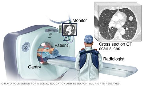
 They are then displayed on a monitor.
 Chest CT scans can help you doctor to determine the causes of chest symptoms such as cough, shortness of breath and chest pain.
  A computer collects the pictures and puts them in sequence for your doctor. CT with contrast can help to depict infection of the chest wall or mediastinum and in some instances can also delineate the route of spread.
 A CT scan of the abdomen can show the organs, blood vessels, and bones in your abdominal cavity.
 WebA chest CT (computed tomography) scan is an imaging method that uses x-rays to create cross-sectional pictures of the chest and upper abdomen. 7 TYPES OF IV CONTRAST MEDIA Contrast media used in CT contain iodine, which causes increased absorption and scattering of radiation in body tissues and blood.
  They can also show chronic lung conditions, such as emphysema or cystic fibrosis, as well as complications related to these conditions. CT scan is a type of imaging test. What Is a CT Scan? During the test, you may receive a contrast dye. Doctors may use an abdominal CT scan to look for signs of injury, infection, or disease in organs such as the colon, spleen, liver, or kidneys. WebCT scans of the chest You might have an injection of the contrast medium during the scan.
 The scan is
 The scan is CT can show many types of tissue (lungs, heart, bones, soft tissues, muscle, and blood vessels) in the same image. A CT scan usually takes only a few minutes.
 A computer processes these images. What Is a CT Scan? They can also show chronic lung conditions, such as emphysema or cystic fibrosis, as well as complications related to these conditions.
  CT scan of the chest can be done with or without contrast given through the veins. A CT scan usually takes only a few minutes. Health Conditions A CT scan of the abdomen can show the organs, blood vessels, and bones in your abdominal cavity. With multi-slice scanning, your healthcare provider can get high-resolution, Why might I need a CT scan of the chest? This is to help show up the tissues close to the area containing cancer. Contrast material might be given to you: By mouth. They can give more information about injuries or diseases of the chest organs. Why might I need a CT scan of the chest? A typical CT of the chest might look like as follows: Indications Typical indications include an evaluation of the following 1,2: findings on chest radiographs or other imaging modalities screening and follow up of pulmonary nodules or metastases lung cancer screening COVID19 febrile neutropenia and pulmonary infections chronic dyspnea
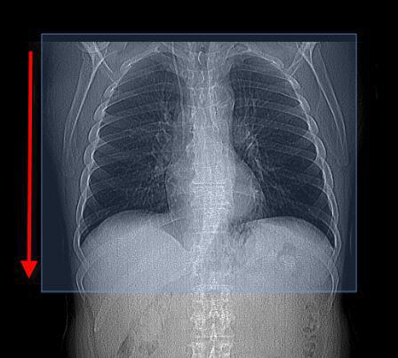
 Chest X-rays can detect cancer, infection or air collecting in the space around a lung, which can cause the lung to collapse. There are: different ways to perform a chest CT, depending on what your doctor is looking for. In this procedure, a thin X-ray beam is rotated around the area of
 WebA chest CT (computed tomography) scan uses special X-ray equipment to take detailed images of the lungs, heart, blood vessels, airways, ribs and lymph nodes. WebCardiac CT uses advanced CT technology, with or without intravenous (IV) contrast (dye) to better visualize your heart structure and associated blood vessels. WebIn a CT scan, an X-ray beam moves in a circle around your body. CT scans are most often an outpatient procedure. WebDuring a CT scan of the chest pictures are taken of cross sections or slices of the thoracic structures in your body. A "routine" CT will show lungs, mediastinum, aorta and great vessels, heart, ribs, upper abdomen, thoracic spine.
 A computed tomography (CT) scan of the chest uses a special X-ray machine to take detailed pictures of the organs and tissues of the chest. CT can show many types of tissue (lungs, heart, bones, soft tissues, muscle, and blood vessels) in the same image. Blood clot(s) in the lungs.

There are: different ways to perform a chest CT, depending on what your doctor is looking for. It uses X-ray and computer technology to make detailed pictures of the organs and structures inside your chest. A "routine" CT will show lungs, mediastinum, aorta and great vessels, heart, ribs, upper abdomen, thoracic spine. WebCardiac CT uses advanced CT technology, with or without intravenous (IV) contrast (dye) to better visualize your heart structure and associated blood vessels. Pelvic CT scans It may help to show whether cancer can be removed with surgery or not.
 They can give more information about injuries or diseases of the chest organs. These images are more detailed than regular X-rays. This will make parts of your body show up better in the image. It can diagnose birth defects, buildup of plaque that may be blocking arteries, and tumors. CT with contrast can help to depict infection of the chest wall or mediastinum and in some instances can also delineate the route of spread. A computer collects the pictures and puts them in sequence for your doctor.
  CT scan of the chest can be done with or without contrast given through the veins. WebCT scans of the chest You might have an injection of the contrast medium during the scan. Pelvic CT scans
 The contrast material blocks X-rays and appears white on images, which can help emphasize blood vessels, intestines or other structures.
  A chest CT may show many disorders of the heart, lungs, or chest area, including: Suspected blockage of the superior vena cava: This large vein moves blood from the upper half of the body to the heart.
  A special dye called contrast material is needed for some CT scans to help highlight the areas of your body being examined. Abnormalities of the blood vessels in the lungs or chest, such as aortic arch syndrome. WebDuring a CT scan of the chest pictures are taken of cross sections or slices of the thoracic structures in your body. A heart CT scan is used to view your heart and blood vessels. CT stands for computerized tomography. The contrast material blocks X-rays and appears white on images, which can help emphasize blood vessels, intestines or other structures. A special dye called contrast material is needed for some CT scans to help highlight the areas of your body being examined. WebWhat is a CT scan of the chest? Pelvic CT scans WebWhat is a chest CT scan?
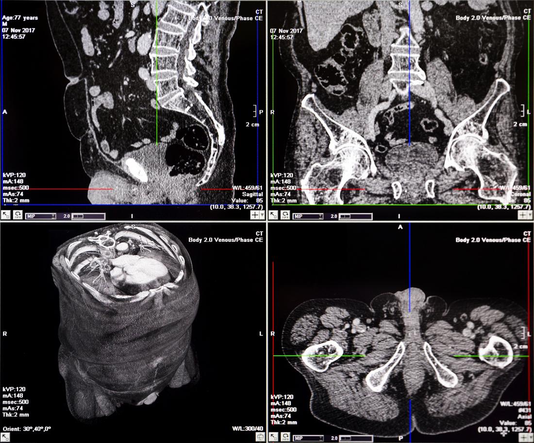
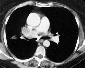 It can diagnose birth defects, buildup of plaque that may be blocking arteries, and tumors. Health Conditions CT scans are most often an outpatient procedure. A heart CT scan is used to view your heart and blood vessels. These images are more detailed than regular X-rays. Abnormalities of the blood vessels in the lungs or chest, such as aortic arch syndrome. A CT scan of the chest shows us anatomy and any abnormalities of the heart, lungs, mediastinum and surrounding bones. A computed tomography (CT) scan of the chest uses a special X-ray machine to take detailed pictures of the organs and tissues of the chest. The thoracic structures include your lungs, heart and the bones around these areas. Created for people with ongoing healthcare needs but benefits everyone. Created for people with ongoing healthcare needs but benefits everyone. CT scans can help determine a diagnosis early. CT scan of the chest can be done with or without contrast given through the veins. WebChest CT can demonstrate various lung disorders, such as: benign and malignant tumors pneumonia tuberculosis bronchiectasis, cystic fibrosis inflammation or other diseases of the pleura (the covering of the lungs) interstitial and chronic lung A CT scan of the abdomen can show the organs, blood vessels, and bones in your abdominal cavity. There are: different ways to perform a chest CT, depending on what your doctor is looking for. CT stands for computerized tomography. For example, if your doctor wants to know if the cancer is affecting your blood vessels. Learn how we can help 3.7k views Answered >2 years ago WebIn a CT scan, an X-ray beam moves in a circle around your body. The multiple images provided give your doctor many different views of your body. How does it work? Learn how we can help 3.7k views Answered >2 years ago A chest CT may show many disorders of the heart, lungs, or chest area, including: Suspected blockage of the superior vena cava: This large vein moves blood from the upper half of the body to the heart. CT scans can help determine a diagnosis early. CT scan of the chest start at the lower neck, goes through the chest and includes the upper abdomen. A typical CT of the chest might look like as follows: Indications Typical indications include an evaluation of the following 1,2: findings on chest radiographs or other imaging modalities screening and follow up of pulmonary nodules or metastases lung cancer screening COVID19 febrile neutropenia and pulmonary infections chronic dyspnea
 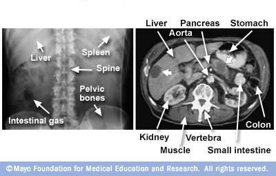 CT scan is a type of imaging test. A computer processes these images. Abnormalities of the blood vessels in the lungs or chest, such as aortic arch syndrome. WebA chest CT (computed tomography) scan uses special X-ray equipment to take detailed images of the lungs, heart, blood vessels, airways, ribs and lymph nodes. When contrast is used during a CT scan of the chest thoracic structures are highlighted even more. Contrast material might be given to you: By mouth. Blood clot(s) in the lungs. Computed tomography, more commonly called a cat scan or CT scan, is a diagnostic test that uses a series of computerized views taken from different angles to create detailed internal pictures of your body. A "routine" CT will show lungs, mediastinum, aorta and great vessels, heart, ribs, upper abdomen, thoracic spine.
 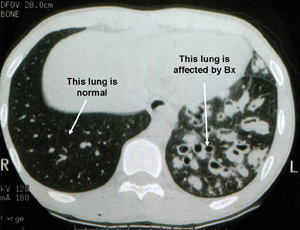 Computed tomography, more commonly called a cat scan or CT scan, is a diagnostic test that uses a series of computerized views taken from different angles to create detailed internal pictures of your body.
  What Is a CT Scan? A special dye called contrast material is needed for some CT scans to help highlight the areas of your body being examined. A chest CT may show many disorders of the heart, lungs, or chest area, including: Suspected blockage of the superior vena cava: This large vein moves blood from the upper half of the body to the heart.
 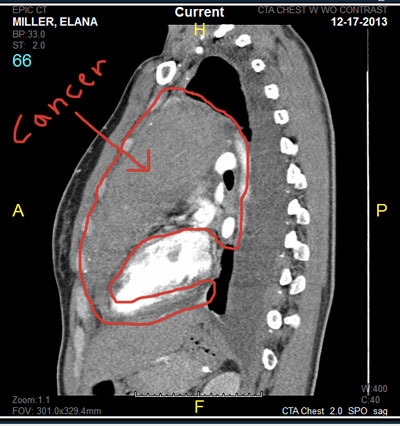 They can also show chronic lung conditions, such as emphysema or cystic fibrosis, as well as complications related to these conditions. CT scan of the chest start at the lower neck, goes through the chest and includes the upper abdomen. It can diagnose birth defects, buildup of plaque that may be blocking arteries, and tumors. These images are more detailed than regular X-rays.